


Dr. Rosalind Franklin and “Photo 51” Provided Valuable DNA Findings
- Susan Stoderl

- Mar 20
- 2 min read
Rosalind Franklin (1920–1958) was an English chemist who used special X-ray images of tiny structures, known as X-ray crystallography. She examined molecules to understand their shape and structure. Her work helped reveal the molecular structures of DNA and RNA, as well as those of viruses, coal, and graphite.
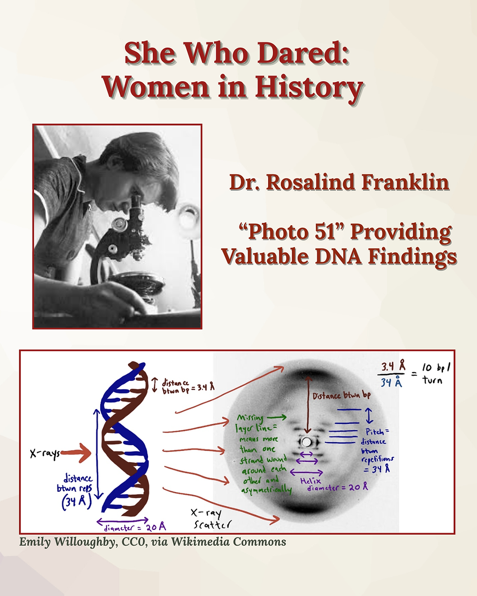
From 1951 to 53, Franklin used X-ray crystallography to study DNA fibers at King’s College London, with her student, Raymond Gosling. They produced exceptionally clear diffraction images, with the most famous being “Photo 51.” The image provided evidence that DNA has a distinctive X-shaped helical structure. She also identified two forms of DNA, A and B. Her insights enabled the construction of an accurate model. Her data and images led to the double helix model published in 1953 by James Watson and Francis Crick. Other related papers by Franklin and colleagues appeared in the same issue of “Nature”.
After leaving King’s College, Franklin established a research group at Birkbeck College, University of London, where she turned her attention to the structure of viruses. She made important advances that helped lay the foundations of structural virology, particularly through studies of helical and spherical viruses.
Rosalind Franklin developed ovarian cancer in 1956 and died in 1958. Some wondered whether all the X-ray work she performed over her lifetime could have played a role in her developing the disease. The one certain thing is that her premature death prevented her from being recognized for her brilliance. Watson, Crick, and Maurice Wilkins received the Nobel Prize in Physiology or Medicine for the discovery of DNA’s structure. Since the Nobel Foundation does not award Nobel Prizes posthumously, Rosalind did not receive what she had equally earned.


Comments